


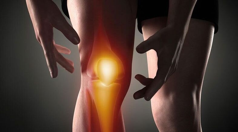
Knee pain- This is a sign of pathological processes that affect the cartilaginous, bony or soft tissue structures of the femoral-tibial and femoral-patellar joints. Arthralgia can be based on trauma, inflammatory and degenerative diseases of the joint apparatus and periarticular structures. Patients may complain of sharp, aching, burning, throbbing, and other types of pain that occur at rest or when moving, leaning, bending, and stretching the leg at the knee. Diagnosis of causal pathology includes instrumental imaging methods (Rg, ultrasound, CT or MRI, arthroscopy), joint capsule puncture, biochemical and immunological analysis. Until the diagnosis is clarified, rest, joint immobilization, NSAIDs and analgesics are recommended.
Causes of knee pain
Traumatic injury
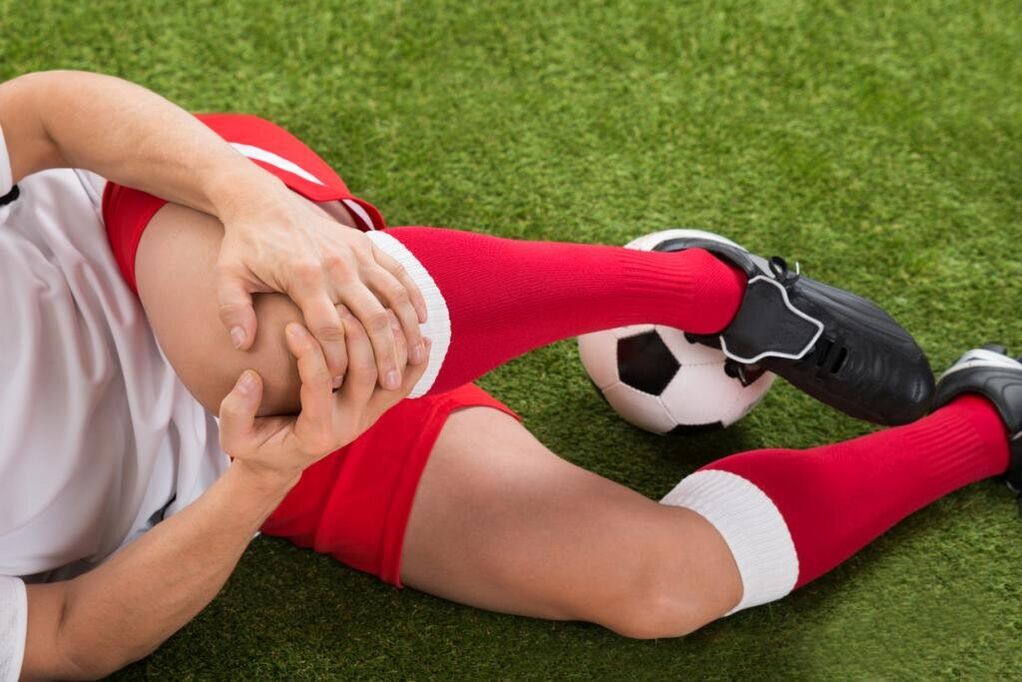
They are usually the result of trauma in the household, they are often found in athletes: runners, jumpers, participants in sports. It develops by falling, hitting or twisting the leg. It is manifested by sharp pain at the time of injury. In the future, the pain syndrome becomes less pronounced, accompanied by an increase in edema. Scratches and bruises are possible. As the frequency increases, the following injuries are identified:
- Knee injury. . . It occurs when falling on the knee or a direct blow to the knee. At the beginning, the pain is sharp, hot, sometimes burning, but bearable, later - dull, painful, intensified by movements. Bruises are possible. The footrest is preserved. Sometimes the knee injury is complicated by hemarthrosis, in such cases the joint gradually increases in volume, becomes spherical, and a feeling of pressure or cracking is added to the pain syndrome.
- Ligament rupture.It is located after twisting the leg, forcibly twisting it, bending it or stretching it excessively in a non-physiological position. Painful sensations are stronger than with a bruise, at the same time with the appearance of pain a person can feel something tearing (similar to ordinary tissue). It is accompanied by significant limitation of movement, support, twisting of the extremities, and rapidly growing hemarthrosis.
- Intraarticular fractures. . . They are detected by bumps, falls and twisting of the leg. In case of injury, the person feels very sharp, often unbearably sharp pain, sometimes a squeak is heard. Patients with an intra-articular fracture themselves describe their feelings as follows: "the pain is such that it darkens in the eyes, the world ceases to exist, you don’t understand anything. " After that, the pain becomes less severe but remains of high intensity. Support is usually impossible, movement is almost completely restricted. Edema and hemarthrosis progress rapidly.
- Dislocation.Is the result of a blow or fall to the knee. At the moment of dislocation of the patella, a sharp pain occurs, followed by a feeling of bending the leg and a shift in the knee. Movement is not possible, the reference function can be saved. A pronounced deformity is visible on the anterior surface of the knee, which is subsequently smoothed out due to increasing edema. Sometimes hemarthrosis joins.
- Pathological fractures.They occur with minor injuries, are the result of reduced bone strength in osteoporosis, osteomyelitis, tuberculosis, bone tumors. The pains are painful, dull, reminiscent of the bruise pain syndrome. Signs that indicate a pathological fracture are limitation or impossibility of support on the leg, feeling of instability in the knee, sometimes deformity, crunching of bones during movement.
- Damage to the meniscus.Meniscus cracks occur during twisting, impact, intense forced bending or extension of the knee, sharp turn with a fixed leg. At the beginning, the person feels a special click and a sharp shooting pain in the depth of the joint. Then the pain decreases somewhat, but it becomes diffuse, sometimes - burning, shooting, intensifies when trying to support and move. Knee volume increases due to edema and hemarthrosis. Support becomes impossible, movements are severely limited.
Inflammatory pathologies
They can be infectious and non-infectious (post-traumatic, toxic-allergic, metabolic, post-vaccination). Abundant blood supply to the synovial membrane and periarticular tissue promotes the rapid development of inflammation in response to direct and indirect effects, and a large number of nerve endings elicit a pronounced pain response. The inflammatory process is often accompanied by synovitis (accumulation of aseptic fluid in the joint), with infection may accumulate pus.
- Arthritis.Gonarthritis occurs after injury, sometimes complicating infectious diseases, is detected in rheumatic diseases. It can be acute or chronic. Knee pain is usually dull, aching, pressing or pulling. At the beginning, the pain is not intense and occasional, it intensifies in the evening or after exercise. Then the initial pains join, the intensity and duration of the pain syndrome increase. The joint swells, the skin over it turns red, his temperature rises. In synovitis, the contours of the knee are smoothed, there is a feeling of cracking. With fertilization, the severity of pain increases sharply, they twitch, deprive of sleep.
- Synovitis.It is not an independent disease, it complicates many acute and chronic pathologies of the joint. It is formed within a few hours or days. At first the pain is slight or absent, a feeling of fullness prevails. The knee is spherical, with a large amount of fluid, the skin is shiny. Movement is somewhat limited. During the infection, the pain becomes pronounced, pulsating, twitching, and intensifies at the slightest movement and touch.
- Bursitis.Inflammation of the joint capsules located in the patella and popliteal fossa usually occurs when the knee is overloaded and its repeated injuries (for example, with constant support on the knees). In bursitis, the pain is local, dull, not intense, it appears at a certain position of the extremities, after a characteristic load, it decreases when the position of the leg changes, massaging the affected area. If the back bag is affected, painful sensations are possible when ascending or descending stairs. Sometimes minor local edema is found. With suppuration of the bursa, the pain becomes sharp, twitching, burning, combined with hyperemia, edema of the affected area, symptoms of general intoxication.
- Tendinitis.It is usually found in obese men and athletes, affecting their own patellar ligament. Initially, the pain syndrome occurs only during very intense exertion, then during standard sports loads, then during daily physical activity or at rest. The pain in tendinitis is localized in front just below the knee, dull, receding, with disease progression, sometimes paroxysmal, in some cases accompanied by mild redness and swelling, increased pressure. Movement is usually complete, rarely slightly restricted. Rupture or rupture of the ligament is possible due to a decrease in its strength.
- Lipoarthritis.Hoff's disease affects the layers of adipose tissue that lie beneath the patella. It is observed with constant overload of the knee or becomes a consequence of an old injury. It affects athletes more often, older women. The person complains of dull aching pains in combination, a certain limitation of extension. With the worsening of the pathology, the pain begins to bother at night, there is a feeling of instability of the knee, bending of the leg. A soft crack or creak is heard when pressing on the side of the patella.
Autoimmune processes
The cause of the disease of this group is the creation of antibodies to normal cells of the organism with the development of immunocomplex aseptic inflammation of the synovial membrane and cartilage, a phenomenon of vasculitis. The pathologies are in most cases chronic, without treatment they are prone to progression, and are often the cause of disability.
- Rheumatoid arthritis.Defeat is usually bilateral. With minimal activity of the autoimmune process, the pain is weak or moderate, intermittent, pulling, pressing, accompanied by morning stiffness. With moderate activity, the patient complains of periodic prolonged pain, pressure or shooting of moderate intensity, not only during movement, but also at rest. There is multiple stiffness, moderate recurrent synovitis. With high activity of rheumatoid arthritis, the pain is strong, diffuse, exhausting, wavy in nature, intensified in the morning. Stiffness becomes constant, a large amount of fluid accumulates in the knees, and contractures are created over time.
- Systemic lupus erythematosus.Arthralgias are often symmetrical, although one joint may be affected. They can occur at any stage of the disease; with a recurrent course of SLE, reminiscent of rheumatoid arthritis. With low activity of the process, the pain is short-lived, non-intense, local, painful, traction. In severe cases, the pain syndrome progresses, the pain is wavy, interferes with night sleep, becomes prolonged, diffuse, intensifies with movement, in combination with synovitis, edema, hyperemia.
- Rheumatism.Joint pain is one of the first manifestations of rheumatic fever, it appears 5-15 days after an acute infection, it affects several joints at once (usually paired). The pains are quite short-lived, but intense, they migrate from one joint to another, they differ in nature from pulling or pressing to burning or pulsing. The knees are swollen, hot, the skin over them reddened. Movement is very limited. After a few days, the intensity of the pain decreases, the movements resume. In some patients, the residual effects in the form of moderate or mild dull pain last for a long time.
- Reactive arthritis.It occurs more often 2-4 weeks after intestinal and urogenital infections, usually affecting one or two joints of the lower extremities, in combination with urethritis, conjunctivitis. Reactive arthritis is preceded by increased urination, pain and burning in the urethra, tearing and cramps in the eyes. Knee pain is strong or moderate, constant, wavy, painful, pulling, twitching, combined with limited movement, worsening of the general condition, fever, severe swelling and redness of the affected area. Painful feelings and signs of inflammation last from 3 months to 1 year and then gradually disappear.
Degenerative-dystrophic processes
They develop as a result of metabolic disorders in the structures of the joint and periarticular soft tissues. They have a chronic course, which progresses over many years. It is often accompanied by the formation of calcifications, cysts and osteophytes, deformation of the knee surface. With significant destruction of joint surfaces, they lead to pronounced impairment of movement and support function, become a cause of disability and require the installation of an endoprosthesis.
- Osteoarthritis.It develops for no reason or in the background of various injuries and diseases, mainly in the elderly and middle-aged people. Initially, the pain is weak, short-lived, usually receding or aching, occurs with prolonged exertion, and disappears at rest, often accompanied by creaking. Gradually, the pain syndrome intensifies, the knees begin to ache "over time", and at night there is a restriction of movement. Characteristics of gonarthrosis are initial pains (pain until you "disperse"), periodic attacks of sharp cutting, burning or shooting pains due to blockade. In periods of exacerbation, synovitis often occurs, in which the pain becomes constant, pressing, shooting.
- Meniscopathy. . . It is usually found in athletes, people whose work involves significant strain on the knee joint. It is manifested by unilateral local deep pain within the knee at the level of the joint space, more often in the outer half of the knee. The pain intensifies during movement and subsides at rest, it can be dull, pressing or pulling. With progression, acute shooting pains occur when trying to move. A small painful formation is sometimes felt on the anterolateral surface of the joint in the projection of pain.
- Tendopathies. . . Tendons near the knee are affected. In the initial phase, they are manifested by short-term local superficial pain at the peak of physical activity. After that, painful sensations occur at moderate and then light loads, limiting normal daily activity. The pain is pulling or painful, directly related to active movements, it is not detected during passive extension and bending of the knee, sometimes accompanied by squeaking or cracking. In the area of the lesion, it is possible to probe the site of greatest pain. Local signs of inflammation (edema, hyperemia, hyperthermia) are insignificant or absent.
- Osteochondropathy.Children and young people get sick more often, the duration of the disease is several years. They usually begin gradually with mild lameness or occasional, non-intense dull pain, increased exertion, which disappears at rest. With the progression of osteochondropathy, the pain becomes strong, constant, pressing, burning or stinging, followed by strong lameness, limited movement and difficult reliance on the extremity. Then the pain gradually decreases, the support function is restored.
- Chondromatosis.It is usually diagnosed in older men, less commonly in infants. Chondromatosis of the joints is manifested by moderate dull pain resembling waves, which often worsen at night and in the morning. Movement is limited, followed by crunch. Sometimes blockages occur, which are characterized by sudden sharp pain, inability or severe restriction of movement. With the development of synovitis, the pain takes on the character of a cracking, combined with an increase in knee volume, soft tissue swelling, and a local increase in temperature.
Tumors and tumor formations
Pain syndrome can be caused by a cyst, benign or malignant tumor that directly affects the joint or periarticular tissue. In addition, knee pain can serve as an alarming signal of hypertrophic arthropathy, paracancrotic polyarthritis - paraneoplastic syndromes characteristic of lung cancer, breast cancer and other oncological processes.
- Baker's cyst.It is a hernial protrusion in the popliteal fossa. In the initial stages, it manifests as uncomfortable feelings or mild local pain along the back of the knee. In the background of an increase in Baker's cyst due to compression of nearby nerves, burning or shooting pains, numbness or tingling in the sole area may occur. The symptoms are worse when you try to bend the knee as much as possible. An elastic, mildly painful tumor-like formation is sometimes felt in the popliteal fossa.
- Benign tumors.It includes chondromas, osteochondromas, non-ossifying fibromas and other neoplasms. They are characterized by a prolonged asymptomatic or low-asymptomatic course, they may be manifested by vague and occasional local non-intense pain. In large neoplasms, a solid formation is felt, sometimes synovitis develops.
- Malignant neoplasms.The most common malignant tumors that affect the joint area are synovial sarcoma, osteosarcoma and chondrosarcoma. They are manifested by dull local vague pains, sometimes with a certain circadian rhythm (up at night). The intensity of pain increases, they become sharp, cut, burn or twitch, spread along the knee and adjacent tissues, accompanied by deformities, edema, synovitis, dilation of the saphenous veins, violation of the general condition, the formation of contracture. Palpation reveals a painful tumor-like formation. When the process starts, the pain is unbearable, unbearable, exhausting, deprives you of sleep and is not eliminated by non-narcotic analgesics.
Invasive operations and manipulations
The pain syndrome is caused by damage to the knee tissue during invasive procedures. The severity of the pain directly depends on the trauma of the manipulation of the knee joint. With the penetration of pathogenic microbes into the joint area, the pain is caused by inflammatory changes.
- Manipulation.The most common procedure is a puncture. The pain after the puncture is short-lived, not intense, subsides quickly, is localized in the projection of the puncture, which is usually performed on the outer surface of the knee. After the biopsy, the pain may initially be twitching, then become dull and disappear after a few days.
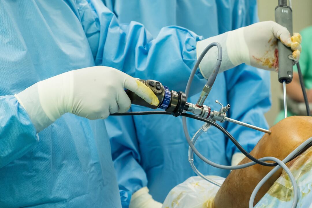
- Operations.After arthroscopy, the pain is moderate, at first quite acute, and then dull, which subsides after a few days or 1-2 weeks. After arthrotomy, the pain syndrome is more intense, it can last up to several weeks due to significant tissue damage. Usually in the first 2 to 3 days after the intervention, patients are prescribed analgesics, then the pain becomes weak and gradually disappears.
Psychosomatic states
Sometimes knee arthralgia occurs in the absence of an organic basis (trauma, inflammation, destruction, etc. ) under the influence of psychological factors. Such pain is believed to play a protective role as it helps reduce emotional stress by turning experiences into physical sensations. The peculiarity of such pains is their indeterminate nature, inconsistency, absence of visible changes, clear connection with physical activity and other objective provoking factors. Meteopathic arthralgias are observed in people who are sensitive to changes in atmospheric pressure.
In addition, radiation of knee pain is possible in coxarthrosis, lumbar osteochondrosis, Perthes' disease, fibromyalgia, sciatic nerve neuropathy. However, in these pathologies, painful syndromes of other localization usually come to the fore. Additional risk factors that increase the likelihood of knee injuries and diseases are overweight, professional sports, hypovitaminosis, metabolic disorders, and old age. Hypothermia, stress, physical exertion, and eating disorders can be provoking factors for worsening chronic pain.
Poll
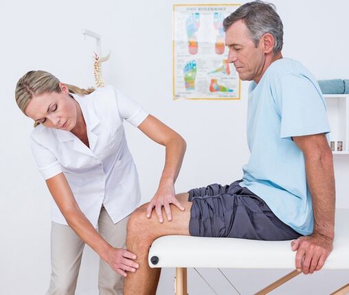
The diagnostic test algorithm is based on taking into account the nature of the pain syndrome, its duration, identification of accompanying symptoms and events that precede the onset of knee pain. During the first visit to the doctor (traumatologist-orthopedist, surgeon, rheumatologist), a visual examination and palpation of the knee is performed, as well as an assessment of the volume of active and passive movements. Taking into account the obtained data, in the future the patient can be assigned:
- Laboratory blood tests. . . Complete blood count helps to detect hematological changes characteristic of acute infectious and inflammatory process (leukocytosis, increased ESR), eosinophilia, typical of an allergic reaction. Biochemical and serological studies are most informative for autoimmune diseases characterized by the production of specific acute phase proteins and immunoglobulins (CRP, rheumatoid factor, ASL-O, CEC, antibodies to DNA, etc. ).
- Radiography.The basic diagnostic method is an X-ray of the knee joint in 2 projections. The presence of pathology is indicated by changes in the contours of the joint head and cavity, narrowing of the joint space, changes in the thickness of the end plates, the presence of marginal defects at the joint ends of the bones, osteolysis and bone destruction. . In some diseases (meniscus trauma, Baker's cyst), contrast arthrography shows the highest sensitivity.
- Arthrosonography. . . Knee ultrasound is a fast, inexpensive, affordable, and highly informative diagnostic method. It allows to assess the presence of effusions and free bodies in the joint cavity, to identify damage and pathological changes in the periarticular soft tissues (signs of calcification, bleeding, etc. ). They help to differentiate the etiology of joint pain with great precision.
- CT and MRI. . . They are the methods of choice for arthropathy of any genesis. They are used for a more detailed assessment of the nature and degree of pathological changes, for the identification of signs typical for traumatic, inflammatory and tumor lesions of bone structures and soft tissues. CT and MRI of the joints are usually used with limited information content from other instrumental studies.
- Joint puncture. . . It is performed when there is an indication of accumulation of exudates or transudates in the joint capsule. As part of the differential diagnosis of inflammatory, degenerative and tumor diseases, a cytological, bacteriological or immunological study of synovial fluid is performed. To diagnose autoimmune damage to the knee joint, tuberculous arthritis, synovioma, it is extremely important to do a biopsy of the synovial membrane.
- Arthroscopy. . . The purpose of invasive endoscopic diagnostics can be to take biopsy samples, to clarify the necessary diagnostic information during the visual examination of the joint elements. In some cases, diagnostic arthroscopy turns into therapeutic (atroscopic removal of intra-articular bodies, meniscectomy, ligament autoplasty, etc. ).
Symptomatic treatment
Treatment of the cause of knee pain is carried out differently, taking into account the identified disease. At the same time, symptomatic care is an essential part of a comprehensive treatment process aimed at reducing discomfort and improving quality of life. Immediately after the injury, it is recommended to apply a cold compress to the knee area - this will help reduce sensitivity to pain. Ethyl chloride has a local cooling and anesthetic effect. In all cases, resting the knee helps reduce pain. It is necessary to limit movement, give the leg a position where pain is minimal. When walking, a fixation bandage is placed on the knee, and immobilization of the extremities with the help of plaster is possible.
In the acute period of injury or illness, it is strictly forbidden to massage the knee, put on warm pads and wear high-heeled shoes. The main classes of drugs used for the symptomatic treatment of pain and inflammation are analgesics and NSAIDs in the form of ointments, tablets and injections. These measures can only temporarily reduce pain, but not eliminate the underlying cause of arthralgia. Therefore, all cases of knee pain require qualified diagnosis and treatment, and some conditions (fractures, dislocations, hemarthrosis) require urgent medical attention. You can not delay a visit to the doctor if the pain is combined with a change in the shape of the knee (swelling, smoothing contours, asymmetry), inability to perform flexion-extensor movements, ballot patella, impaired limb support.